RESEARCH
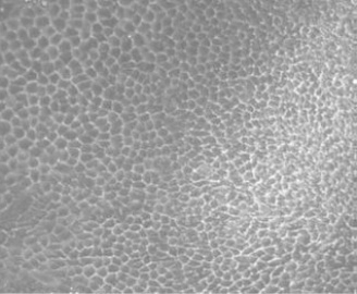
Phase contrast image of RPE cell
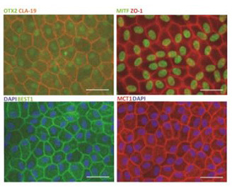
AhRPE cells isolated from human cadaveric tissue (DIV 30), fixed and immunostained for RPE markers (Scale bar = 20μm.)
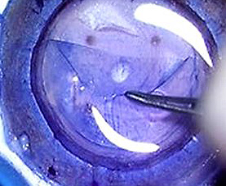
Cornea processing-dissection of
Descemet membrane and endothelium


The Eye-Bank's Ocular Tissue Research Program
The Eye-Bank provides quality donor eye tissue for research into treatments and cures for other eye diseases. Control and diseased ocular research tissues are available to assist with ocular, neurodegenerative and metabolic studies.
Fully Equipped Ocular Laboratory
- Biosafety cabinet 2 (BSC2)
- Dissection microscope;
- Light microscope with camera;
- CO2 incubator
- Konan CellCheck D+ specular microscope
- Slit lamp biomicroscope
- Zeiss Cirrus 5000 OCT machine
- Lumedica OCT machine
Research Tissue Available
Tissues
- Whole eye
- Posterior pole
- Iris
- Lens
- Retina
- Conjunctiva
Primary adult ocular cells*
- Adult human primary retina pigmented epithelial cells (ahRPE-sc)
- Adult human primary Müller cells
Tissue processing*
- RNA, DNA and Protein extraction
- Dissection epithelium or endothelium layer of cornea
* Customization available upon request
Preservation Methods
Fixatives*
- Formalin / formaldehyde
- Davidson's fixative
- Hartmann's fixative
- RNAlater
- 8M Urea
- Paraformaldehyde (PFA)
Media*
- Optisol-GS
- DMEM
- Hybernation media
Other*
- Moist chamber
- Flash freezing (LN2)
- Freezing in OCT (optimal cutting temperature) compound
* Customization available upon request